
Why Is It Called a 12-Lead ECG? (Simple Explanation)
A 12-lead ECG gets its name because it records 12 different electrical perspectives of the heart, even though only 10 electrodes are attached to the body. These electrodes detect electrical signals from different angles, and the ECG machine combines these signals to create 12 diagnostic “leads,” or views, of cardiac activity.
Electrodes vs. Leads: What’s the Difference?
One of the biggest misconceptions is that leads are wires.
They’re not.
-
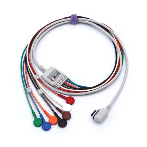
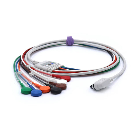
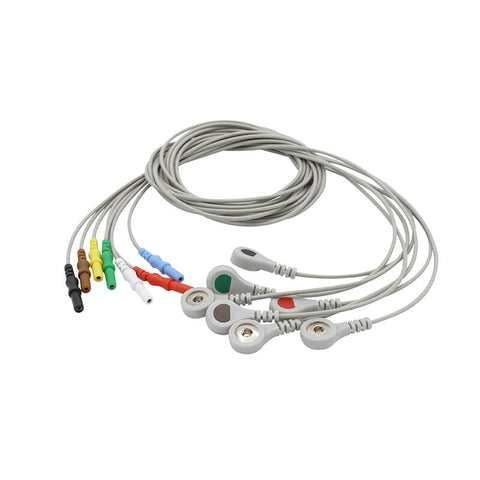
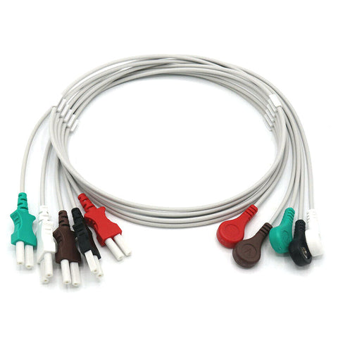
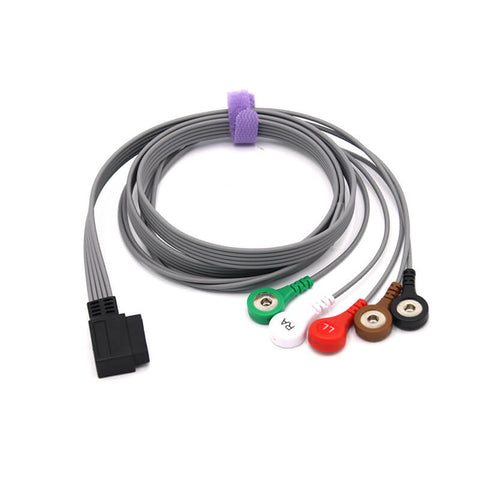
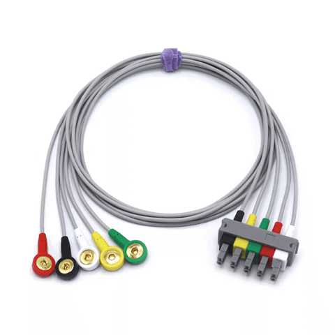
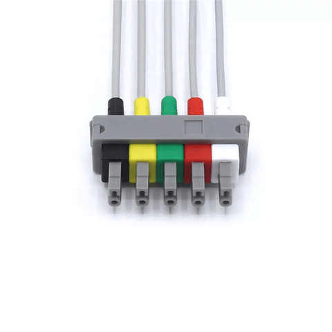
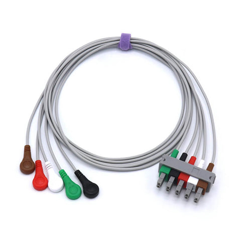
Electrodes = the sticky patches placed on the skin
-
Leads = the electrical views created by comparing signals from different electrodes
So, 10 electrodes can generate 12 unique leads because the ECG machine calculates multiple voltage differences between them.
How the 10 Electrodes Create 12 Leads
1. Limb Electrodes (4 total)
Placed on both arms and legs.
These produce 6 limb leads, giving a vertical-plane view of the heart:
-
Lead I
-
Lead II
-
Lead III
-
aVR
-
aVL
-
aVF
2. Chest Electrodes (6 total)
Placed across the chest (V1–V6).
These generate 6 precordial leads, providing a horizontal-plane view:
-
V1
-
V2
-
V3
-
V4
-
V5
-
V6
Why 12 Leads Matter
By combining all 12 angles:
-
Doctors get a complete 3D electrical picture of the heart
-
Subtle abnormalities such as ischemia, infarction, or conduction blocks become easier to detect
-
It remains the gold standard in cardiac diagnostics
In short, the “12-lead” name reflects the 12 diagnostic views, not the number of electrodes used.