
Why Use a 5-Lead ECG?
Understanding 5-Lead ECG Monitoring and Its Clinical Importance
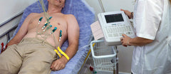
A 5-lead electrocardiogram (ECG or EKG) is one of the most reliable methods for continuous cardiac monitoring, especially in hospital and emergency settings. Unlike a traditional 12-lead ECG, which captures a brief snapshot of the heart’s electrical activity, the 5-lead ECG system allows real-time observation of the heart rhythm, making it invaluable for detecting arrhythmias, ischemic changes, and assessing the effectiveness of cardiac treatments or implanted devices.
In modern healthcare environments—such as intensive care units (ICUs), operating rooms, and ambulance transport systems—the 5-lead ECG has become the standard for ongoing cardiac assessment. It combines accuracy, efficiency, and patient comfort, allowing medical teams to identify subtle cardiac abnormalities before they develop into life-threatening events.
The Primary Purpose of a 5-Lead ECG
1. Continuous Cardiac Monitoring
A 5-lead ECG provides continuous, real-time tracking of a patient’s heart rate and rhythm. This makes it ideal for monitoring patients under anesthesia, in critical care, or during transport. Continuous monitoring allows clinicians to observe moment-to-moment changes in the heart’s electrical activity and respond quickly to emerging issues.
2. Arrhythmia Detection and Tracking
One of the most valuable uses of a 5-lead ECG is its ability to detect and monitor arrhythmias, such as atrial fibrillation, tachycardia, or bradycardia. These rhythm disturbances can appear intermittently, and a single 12-lead ECG might miss them. Continuous 5-lead telemetry helps healthcare providers identify transient arrhythmias and evaluate whether medications or interventions are effective in controlling abnormal rhythms.
3. Ischemia and Myocardial Infarction (Heart Attack) Detection
The 5-lead configuration can also detect early signs of myocardial ischemia—a reduction in blood flow to the heart muscle. By monitoring specific leads, clinicians can identify ST-segment changes that signal potential heart damage or a prior myocardial infarction (MI). This early detection allows for faster diagnosis and intervention, improving patient outcomes.
4. Evaluating Treatment Effectiveness and Device Function
A 5-lead ECG plays a vital role in assessing treatment efficacy, especially for patients with pacemakers, defibrillators, or antiarrhythmic therapy. It helps clinicians confirm that the electrical impulses generated by an implanted device are functioning correctly and that medical treatments are producing the desired cardiac responses.
5. Use in Emergency and Critical Care Environments
The 5-lead ECG is a standard monitoring tool in emergency rooms, intensive care units, operating theaters, and ambulances. Because it provides a balance between simplicity and comprehensive data, it’s ideal for unstable or high-risk patients who need constant observation. Paramedics and emergency medical technicians often use portable 5-lead ECG systems for pre-hospital monitoring, transmitting real-time data to hospitals before arrival.
How a 5-Lead ECG System Works
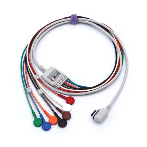
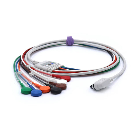
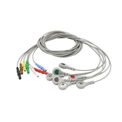
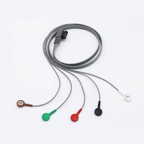
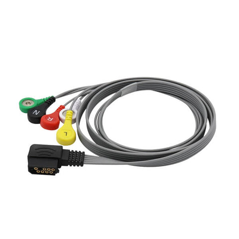
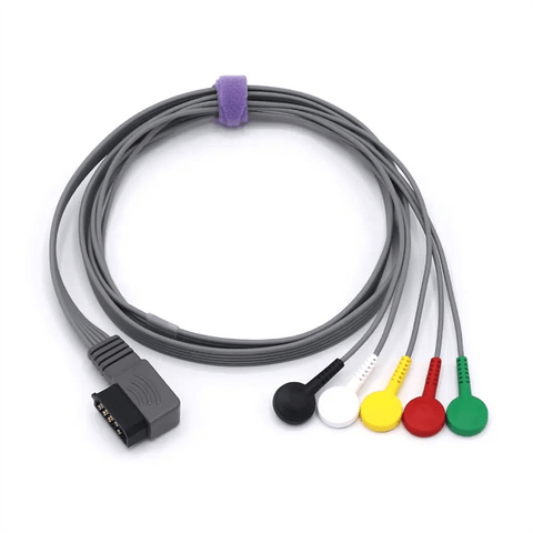
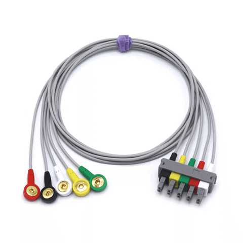
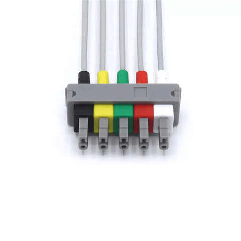
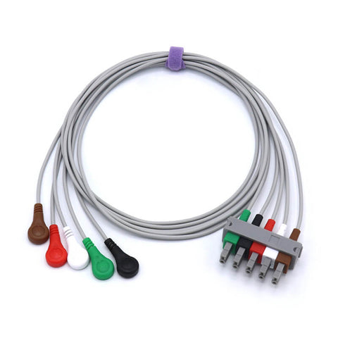
Electrodes and Leads
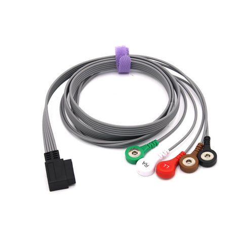
The system uses five electrodes, which are small, adhesive patches placed on the patient’s skin. These electrodes detect the heart’s electrical signals and transmit them through five leads (wires) connected to an ECG monitor.
Standard Placement
In a 5-lead ECG setup:
-
Four limb electrodes are placed on the torso, representing the right arm (RA), left arm (LA), right leg (RL), and left leg (LL).
-
The fifth electrode (V lead) is placed on the chest, typically in the V1 or V5 position, depending on the area of the heart clinicians want to monitor.
This configuration allows clinicians to observe the heart from multiple electrical perspectives, offering better diagnostic precision than simpler three-lead systems.
Data Collection and Visualization
Once the electrodes are in place, the ECG machine records the electrical impulses that cause the heart to contract and relax. The resulting data is displayed as waveforms on a screen or printed on paper. Clinicians can analyze these waveforms to detect abnormal rhythms, conduction delays, or ischemic changes.
Comprehensive Cardiac Views
The 5-lead ECG provides enhanced spatial coverage of the heart’s electrical field compared to a 3-lead ECG. This means clinicians can visualize P-waves, QRS complexes, and ST-segments more accurately—crucial for spotting ischemia or early infarction signs that might otherwise go unnoticed.
Advantages of a 5-Lead ECG Over Other Systems
-
Better Diagnostic Coverage: Offers more data points and lead views than a 3-lead system, improving rhythm and ischemia detection.
-
Continuous Real-Time Data: Enables early identification of cardiac deterioration and immediate response.
-
High Clinical Flexibility: Suitable for both in-hospital and pre-hospital care, from ICUs to ambulances.
-
Compact and Efficient: Balances the comprehensive data of a 12-lead ECG with the simplicity of fewer electrodes, minimizing patient discomfort.
-
Enhanced Patient Safety: Provides round-the-clock monitoring for patients recovering from cardiac surgery, trauma, or those on life-support systems.
In Summary
A 5-lead ECG is a cornerstone of modern cardiac monitoring. It bridges the gap between comprehensive diagnostics and practical, continuous observation, giving clinicians a powerful tool to track a patient’s heart health over time. From detecting arrhythmias and ischemia to verifying treatment success, it plays a crucial role in both acute care and long-term cardiac management.
Whether in an ICU, ambulance, or recovery room, the 5-lead ECG ensures that every heartbeat is monitored, recorded, and understood, supporting faster decisions and safer outcomes for patients.